Choosing Mandibular Reconstruction Plates requires more than matching a plate to a bone defect. In practice, surgeons assess computed tomography scans, occlusion, defect length, remaining bone, and soft-tissue condition. They also consider infection risk, bone quality, and the patient’s healing capacity. These factors can change the preferred design, thickness, and fixation method. A plate that appears strong on paper may still be unsuitable for a thin mandibular segment or limited screw purchase. No plate is perfect.
This guide examines the practical factors behind reliable plate selection. It discusses titanium materials, load-bearing and load-sharing designs, locking screws, plate profiles, and intraoperative contouring. Screw trajectory matters, especially near the inferior border, dental roots, and planned bone grafts. Digital planning can improve accuracy, but surgical findings may differ from the virtual model. Experienced teams should compare manufacturer specifications, peer-reviewed evidence, and institutional protocols before making a decision. Evidence quality is not always consistent. That deserves honest attention.
The most suitable plate depends on the defect, reconstruction method, and patient-specific risks. Important questions include whether vascularized bone is planned, whether radiotherapy is expected, and how much native bone remains. Clear documentation supports follow-up and helps clinicians monitor loosening, breakage, infection, or malocclusion. The smallest detail matters. This article provides educational guidance, not a substitute for clinical judgment. Final decisions should involve qualified oral and maxillofacial surgeons, reconstructive specialists, and the wider care team. Careful collaboration usually produces safer, more defensible treatment planning.
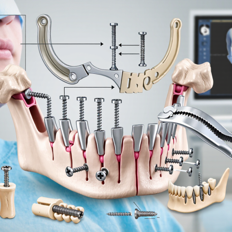
Mandibular reconstruction plates vary in type, geometry, and mechanical purpose. Load-bearing plates bridge large defects when bone continuity is lost. Load-sharing plates support fractures when healthy bone still carries part of the bite force. Choosing between them requires imaging, defect size, bone quality, and the planned graft. Fit matters.
Straight plates suit relatively simple contours, while pre-contoured plates follow the mandibular angle, body, or symphysis more closely. Curved designs can reduce bending during placement. Their holes may be locking or non-locking. Locking holes maintain screw-plate stability, even when the plate does not press tightly against the bone. Non-locking holes allow compression but demand more accurate contact and contouring. Both systems have limitations.
Structural strength depends on thickness, width, hole spacing, and the transition between solid and perforated areas. Wide plates resist bending, but they may increase soft-tissue prominence. Thicker plates can tolerate greater loads, yet they may be harder to contour without distortion. Smooth edges may reduce irritation around the covering tissues. Screw trajectories also matter, especially near tooth roots, the inferior alveolar canal, or a graft interface. Small contour errors can create large stresses. That detail matters.
In practical planning, surgeons compare three-dimensional imaging with the occlusion and remaining bone. A plate that looks strong on paper may perform poorly after repeated chewing forces. A very flexible plate may adapt easily but lose stability. No design is perfect. Final selection should follow patient-specific assessment by a qualified maxillofacial team, with attention to fixation strategy, healing conditions, and follow-up imaging.
How to Choose Mandibular Reconstruction Plates?
Patient and defect factors should drive mandibular plate selection, not habit or availability. GLOBOCAN 2022 estimated 389,846 new lip and oral cavity cancer cases worldwide. Many patients therefore need reconstruction after tumor removal, not simple fracture fixation. Segment length, defect location, bone continuity, and soft-tissue loss must be assessed together. A lateral segment may tolerate different forces than a symphysis defect. It also needs careful contouring.
Load-bearing plates are often considered when the bony bridge is absent or healing is uncertain. Load-sharing designs may suit patients with strong bone contact and stable graft support. Patient factors matter equally. Previous radiation, smoking, diabetes, malnutrition, poor bone quality, and limited oral hygiene can increase wound and hardware complications. Age alone is not a reliable selection rule. It is only one piece.
A 2021 systematic review of vascularized fibula reconstructions reported plate exposure rates near 20%, although results varied widely between studies. That number should encourage caution, not fear. Screw distribution, plate position, tissue coverage, and postoperative care can change the risk. Three-dimensional planning improves accuracy, but it cannot predict every biological response. A tidy algorithm is tempting. Real patients rarely follow one. I would reassess the plan when soft tissue is thin, radiation is extensive, or the defect crosses the midline. Intraoperative findings may still justify a stronger or differently contoured plate.
How to Choose Mandibular Reconstruction Plates?
Material, strength, and design should match the defect, bone quality, and surgical plan. Titanium is widely selected because it is light, corrosion resistant, and generally compatible with imaging. Stainless steel can offer high strength, but its weight and imaging behavior may affect decisions. Material choice is not purely technical. Allergies, previous operations, radiation exposure, and soft-tissue coverage also matter.
Strength must withstand chewing forces without creating excessive stress at the fixation sites. A thicker plate may resist bending, yet it can irritate thin soft tissue. A very rigid construct may also transfer force poorly to weakened bone. Design details deserve close inspection. Locking holes can preserve alignment when bone purchase is limited. Broad plates may spread load, while narrow plates can fit restricted anatomy. Pre-contoured shapes save operating time, but they may not match every jaw. That limitation is easy to underestimate.
Tips: Review three-dimensional imaging before selecting the plate. Check the planned osteotomy, remaining bone, and screw paths. Measure the soft-tissue envelope, not only the defect. Ask whether the plate can be contoured without weakening it. Small gaps around the plate can collect fluid and complicate healing. In practice, the “strongest” option is not always the safest. Surgeon experience, validated mechanical data, and careful intraoperative assessment should guide the final choice. Mistakes happen when design convenience replaces patient-specific planning.
| Evaluation Dimension | Option / Design Feature | Relevant Facts and Typical Values | Clinical or Engineering Implication |
|---|---|---|---|
| Material | Commercially pure titanium | Density is approximately 4.5 g/cm³. Elastic modulus is about 100–110 GPa, substantially closer to cortical bone than stainless steel. | Lightweight and highly corrosion-resistant. The lower stiffness may help reduce, but does not eliminate, stress shielding. |
| Titanium alloy, such as Ti-6Al-4V | Density is approximately 4.4–4.5 g/cm³; elastic modulus is commonly around 105–115 GPa. Yield strength is often approximately 800–950 MPa, depending on grade and processing. | High strength-to-weight ratio and good fatigue performance make it widely suitable for load-bearing mandibular reconstruction plates. | |
| 316L stainless steel | Density is approximately 8.0 g/cm³ and elastic modulus is about 190–200 GPa. Mechanical properties vary considerably with temper and manufacturing method. | Strong and familiar in surgical applications, but heavier and stiffer than titanium. Verify corrosion resistance, imaging requirements, and patient sensitivity considerations. | |
| Strength and Load Demand | Load-bearing reconstruction | Required when the mandible has a critical-size defect, segmental resection, poor bone contact, or delayed graft incorporation. | Select a plate system with adequate bending strength, fatigue resistance, screw fixation, and validated performance for the intended defect pattern. |
| Load-sharing fixation | Used when stable native bone or a well-fitting graft can share functional loads with the plate. | May permit a lower-profile construct, but fixation still depends on bone quality, defect geometry, occlusion, and the number and distribution of screws. | |
| Plate Geometry | Thickness and cross-sectional profile | Common reconstruction plate thicknesses are approximately 2.0–3.5 mm, but the available range depends on the system and indication. | Greater thickness generally increases bending resistance but may increase prominence, soft-tissue irritation, and contouring difficulty. |
| Plate width and edge profile | Wider plates can distribute forces over a larger area. Rounded or beveled edges reduce sharp transitions at the soft-tissue interface. | Balance mechanical stability with available bone surface, soft-tissue coverage, and the risk of plate palpability or exposure. | |
| Precontoured versus manually contoured | Precontoured plates are shaped to an anatomical reference; manually contoured plates can be adapted intraoperatively. | A close fit can reduce intraoperative bending and preserve plate strength. Excessive repeated bending may create local stress concentrations or weaken the construct. | |
| Screw and Hole Design | Locking screw compatibility | Locking screws engage the plate hole and can form a fixed-angle construct; non-locking screws compress the plate against bone. | Locking fixation can be useful in compromised bone or grafted segments because stability is less dependent on plate-to-bone compression. |
| Hole spacing and screw distribution | Biomechanical stability depends on screw number, bicortical or monocortical purchase, distance from the defect, and the quality of the remaining bone. | Distribute fixation on both sides of the defect whenever anatomy permits, while avoiding tooth roots, the inferior alveolar canal, and planned osteotomy or graft zones. | |
| Variable-angle holes | Permit some control over screw trajectory within a manufacturer-defined angular range. | May improve fixation in irregular anatomy, but the approved angle limits and compatible screw types must be followed. | |
| Biocompatibility and Imaging | Corrosion and tissue compatibility | Titanium and titanium alloys form a stable passive oxide layer and are generally considered highly corrosion-resistant in the body. | Material selection should consider allergy history, prior implants, infection risk, and the complete implant system rather than the plate alone. |
| CT and MRI considerations | Titanium generally produces less CT artifact than stainless steel, although artifact depends on geometry, scan parameters, and adjacent materials. Titanium is non-ferromagnetic but may still cause MRI susceptibility artifact. | Review the implant’s verified MRI conditions and anticipate imaging artifacts when postoperative surveillance or radiotherapy planning is important. | |
| Clinical Environment | Soft-tissue coverage | Thin or irradiated soft tissue, intraoral exposure, and limited vascularity increase the risk of wound complications and hardware exposure. | Favor an appropriately low-profile, smoothly contoured construct and ensure adequate vascularized soft-tissue coverage when clinically indicated. |
| Infection and patient factors | Smoking, diabetes, malnutrition, previous radiotherapy, poor oral hygiene, and active infection can impair healing and increase failure risk. | Plate selection cannot compensate for inadequate debridement, unstable occlusion, poor fixation, or insufficient biological support. | |
| Manufacturing and Verification | Additive-manufactured patient-specific plate | Designed from patient CT data and typically manufactured from a medical-grade metal such as titanium alloy. Accuracy depends on imaging, segmentation, design, and production controls. | Can improve anatomical fit and surgical planning, but requires validated design review, manufacturing quality control, and regulatory compliance. |
| Sterilization and system compatibility | The plate, screws, instruments, and sterilization method must be compatible with the device instructions and applicable medical-device standards. | Confirm material grade, screw compatibility, approved bending limits, sterilization instructions, and MRI labeling before use. |
Note: Numerical values are representative ranges for commonly used implant materials and designs. Actual performance depends on alloy grade, heat treatment, geometry, manufacturing process, fixation technique, bone quality, and patient-specific loading.
Mandibular reconstruction plates should match the surgical technique, not merely the defect’s outline. A load-bearing plate may suit a segmental gap with limited bone contact. A load-sharing design can work when healthy fragments meet securely. The distinction matters during chewing, graft healing, and early movement. Plan from computed tomography images, occlusion, and the expected soft-tissue envelope. Do not rely on plate shape alone.
Fixation method also changes the choice. Locking screws can maintain stability when bone quality is reduced or access is limited. Conventional screws may provide compression when fragments can be accurately adapted. Bicortical fixation often improves purchase, but it may increase risk near teeth, nerves, or vessels. Screw length, hole position, and insertion angle need direct review. Small errors become expensive here.
In the operating room, contouring should preserve plate strength and avoid sharp edges beneath thin tissue. A plate that fits a digital model may still conflict with a graft, tracheostomy, or incision. Clinical teams often find that rehearsal improves speed, yet rehearsal can create false confidence. Reassess stability after occlusion is restored and after every critical screw is seated. Document the reasoning, including compromises, because the ideal fixation plan rarely survives unchanged.
Evaluation Criteria for Safety, Function, and Long-Term Outcomes
Plate selection should begin with the patient’s anatomy, not the catalog. In practice, surgeons compare defect length, bone quality, occlusal forces, and soft-tissue coverage. A 2022 systematic review in Oral Surgery, Oral Medicine, Oral Pathology and Oral Radiology reported plate-related complications commonly ranging from 10% to 30% across mandibular reconstruction cohorts. These complications included exposure, loosening, fracture, and infection. The figure varies widely. That matters.
Safety depends on accurate three-dimensional adaptation and stable screw purchase. Computed tomography planning can reduce bending errors, but it cannot replace intraoperative judgment. Evaluate screw trajectory near the inferior alveolar canal. Check plate thickness against functional loading. Consider fatigue resistance when the graft may unite slowly. Long-term function also includes speech, chewing, facial symmetry, and the patient’s ability to maintain oral hygiene. A 2023 systematic review in the Journal of Cranio-Maxillofacial Surgery linked poor soft-tissue coverage and smoking history with higher hardware complications. Evidence remains uneven. Some studies use small samples.
Tips: Request at least three years of follow-up data when comparing plate systems. Review fracture and exposure rates separately. Confirm whether outcomes include irradiated patients. Ask how many screws remain stable at twelve months. Do not judge success from radiographs alone. A comfortable bite may reveal more than a perfect image. Reassess the plan when vascularity, occlusion, or patient compliance changes.
It stabilizes the jaw when bone continuity is lost or healing may be uncertain.
Load-bearing plates carry much of the chewing force across a missing bone segment.
Segment length, defect location, bone continuity, and soft-tissue loss all influence the choice.
Pre-contoured plates follow common jaw shapes and may reduce operating time.
Locking holes stabilize screws within the plate, even without tight bone contact.
Previous radiation, smoking, diabetes, poor nutrition, weak bone, and limited oral hygiene may increase risks.
Wider and thicker plates usually resist bending better.
Titanium is light, corrosion resistant, and generally compatible with imaging.
They should examine three-dimensional imaging, occlusion, remaining bone, soft-tissue coverage, and planned screw paths.
No. It improves accuracy but cannot predict every biological response.
Choosing Mandibular Reconstruction Plates requires a careful balance between anatomical demands, surgical goals, and long-term patient needs. Different plate types and structural designs offer varying levels of contourability, rigidity, and support, so selection should reflect the size, location, and complexity of the mandibular defect. Patient-specific factors, including bone quality, soft-tissue condition, functional expectations, and overall health, also influence the most appropriate solution.
Material strength, fatigue resistance, profile, and adaptability are essential design considerations. The plate should be compatible with the planned surgical technique, fixation method, grafting approach, and available imaging or shaping procedures. A complete evaluation should consider placement accuracy, stability, protection of surrounding tissues, occlusion, speech, chewing function, and ease of follow-up. By assessing these factors together, clinicians can select Mandibular Reconstruction Plates that support reliable healing, reduce complications, and contribute to stable functional and aesthetic outcomes over time.
Virelox